The skin

Build-up of the skin
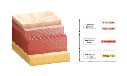
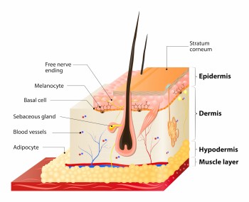
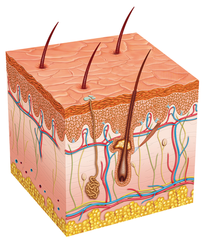
Our skin consists of three layers: the epidermis (outer layer), the dermis (middle layer) and the subcutaneous tissue (bottom layer).
The outer skin (epidermis)
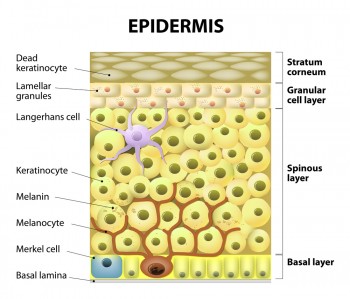
This is the outer layer of the skin. The epidermis itself has several distinct layers as well: basal cell layer (stratum basale), prickle cell layer (stratum spinosum), granule cell layer (stratum granulosum), clear layer (stratum lucidum) and the horned layer (stratum corneum).
Basal layer (stratum basale)
The bottom layer is called the basal layer (stratum basale). The word stratum basale is derived from Latin: stratum means “layer” and basal is “basic” or “ground”. The cells of the basal layer are attached to the basilar membrane (a thin tissue), which separates the epidermis from the dermis.
The cells are produced at the basal layer of the epidermis. They are called keratinocytes, because they form keratin, also known as horn dust. There is constant proliferation of cells in the basal layer of the epidermis.
Cells multiply by partition. The cells produced by this layer, move upwards to the outer layers of the skin, where they continue to grow. Pigment is produced at the basal layer of the epidermis as well.
Prickle cell layer (stratum spinosum)
The layer under the basal layer is the prickle cell layer, also known as stratum spinosum. Spino means “thorn”. Keratinosomes are small cavities surrounded by a membrane (odland bodies). There is a fatlike substance in these odland bodies in the shape of disc-shaped double membrane lipids. Lipids are fats or fatlike substances.
Granule cell layer (stratum granulosum)
The layer on the prickle cell layer is called granule cell layer or stratum granulosum. Granula means “grain”. In this layer the keratinisation (the conversion in horny tissue) takes place. This layer owes its name to its granular look, caused by the keratohyalin granules, a mixture of different kinds of proteins.
Clear layer (stratum lucidum)
The clear layer or the stratum lucidum – which is highly reflective – lies between the layers called the stratum corneum and the stratum granulosum. This layer is not very clear in all places. The stratum lucidum is present only where the skin is especially thick, such as the soles of the feet and the palms of the hands. The cells are flat and lie close to each other. They do not contain any noticeable boundaries.
Horned layer (stratum corneum)
Stratum corneum (corneum means “horny skin”) is the outermost of the five layers of the epidermis and is to form a barrier to protect underlying tissue from infections and prevents dehydration of the skin.
The epidermal lipids (liposomes) lie between the horny cells (corneocytes).Towards the surface the horny layer of the skin becomes increasingly fragile and the spaces between the cells become increasingly larger. The individual cells split apart from each other, loosen and are sloughed off unnoticed as scales when you put on and take off clothes, when you scratch yourself or during an exfoliation treatment for instance. This unperceived, continuous process is called desquamation.
An adult human sheds approximately 10 grams of skin scales a day. This is no reason to worry, since new layers are continuously formed in the basal layer by cell division. The skin renewal process takes about 30 days. The skin of younger people renews slightly faster than the skin of older people.
In psoriasis, the cornification process is also faster. The division takes place more quickly in the basal layer and the relocation of horny cells takes place more quickly. Sometimes this process only takes a week or shorter instead of a month.
Function
Though the epidermis is very thin (in some places less than a tenth of a millimetre), this layer is very important for our protection. The keratinocytes are located so close to each other, that they provide a buffer against external influences. This dense structure also prevents viruses, fungi and bacteria from entering the body, but also protects our bodies from evaporation.
The skin’s ability to retain moisture is mainly due to the composition of the barrier lipids (fats) in the horned layer. The protein structure of the horny cells is important for the moisture-retaining ability as well. These substances, which occur naturally in the body and retain water in the horny layer, are called natural moisturising factors.
Thickened horned layer
In the ordinary course of events, the horny layer (stratum corneum) is a very thin membrane. On places where the skin experiences a lot of pressure, under the sole of one’s for instance, the heel or in the hands, the horny layer thickens. We call this callous. The continuous pressure is a signal for the skin to produce more keratinocytes and to slow down the shedding process. Because of this a thicker horny layer is formed. If it concerns a small and long-lasting pressure point, there is a chance that the horny layer not only thickens, but also ‘grows’ inwards. In this case, acorn is formed. You can have callous and corns removed by a chiropodist.
Melanocytes and Langerhans cells
Between the keratinocytes and the basal layer, there are other cells as well: the melanocytes and the Langerhans cells. Melanocytes are pigment cells producing pigment grains. The pigment of these grains is called melanin. The melanocytes transfer the pigment grains to the keratinocytes via sprouts, giving them a colour. The melanin is not only responsible for the skin colour, but is also a protective layer against the fading effects of the sun’s UV rays. Sometimes UV rays produce too much or too little pigment. This can result in pigmentation disorders.
The Langerhans cells are derived from cells originating in bone marrow. If infection germs or chemical substances enter the body they mobilise the immune system. Langerhans cells are, as it were, cells that keep watch and set off an alarm when there are intruders.
There are no small blood vessels in the epidermis!
The dermis
The largest part of the dermis consists of elastic, nearly inelastic collagen fibres, which are essential to keep skin resilient and firm.
The dermis contains:
- Blood vessels (food and oxygen supply). The blood vessels are also responsible for controlling the body temperature;
- Lymphatic (drainage of waste);
- Nerves (sense of touch, pain conduction, temperature sensitivity);
- Sweat glands to keep your body temperature stable;
- Sebaceous glands that ensure that the skin does not dehydrate.
The subcutaneous fatty tissue (subcutis)
The subcutaneous fatty tissue, also called subcutis, consists of:
- Loose connective tissue with fat cells (adipocytes) in big cushion-like clusters; eighty per cent of the volume of these cells consists of fats (triglycerides);
- Regular dense connective tissues;
- Blood vessels running through the subcutaneous connective tissue.
The fat has three main tasks:
- Energy reservoir;
- Isolation;
- Support function.
Subcutis separates the skin from muscles and tendons in the body. This layer also determines the skin type.
The structure of the skin layer changes with age. The skin layers clearly become thinner. Moreover, the fatty tissues and the elastic fibres decrease. The blood flow declines with and the number of sweat glands decrease, as a result of which the oxygen and nutrient supply declines. All these changes cause the skin to become thin, dry and less elastic with age. The skin becomes more prone to damage and recovers more slowly.
Source: https://www.goingorganic.nl/index.php/Opbouw-van-de-Huid.html